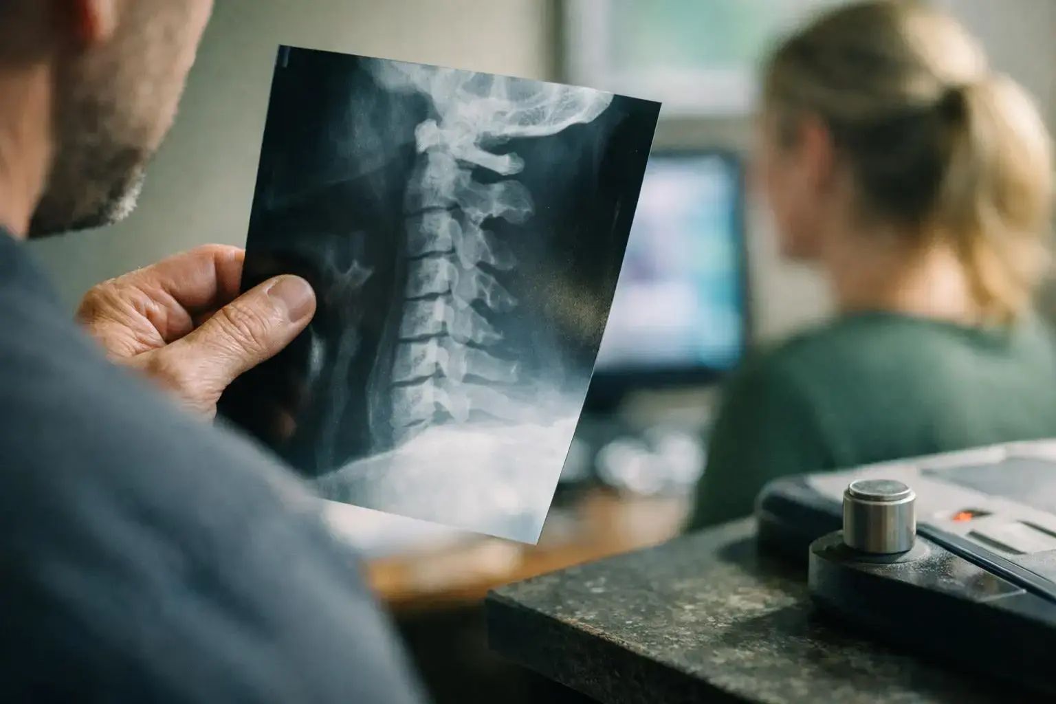
X-Ray is x-Rays are a type of medical imaging that uses safe, invisible electromagnetic waves to create pictures of the inside of the body, especially bones. X-Rays help chiropractors and doctors see fractures, misalignments. Or other problems without surgery. The images show dense structures like bones in white and softer tissues in shades of gray.
Category
Medical imaging
Used for
Viewing bones, joints. And some internal organs
Common confusion
X-Rays show bones clearly but not soft tissues like muscles
Also called
Radiograph, Röntgen ray
Often discussed with
Chiropractic Consultation and Diagnosis, Car Accident Injury Rehabilitation

X-Rays help doctors see inside your body. They use a small amount of radiation. This radiation goes through your body.
Related glossary terms: Chiropractic Adjustment, Cervical Spine, Lumbar Spine.
Bones block most of the radiation. They look white on the image. Muscles and organs let more radiation pass. They look gray.
This helps doctors find problems. They can see broken bones or infections. They can spot misalignments too. No surgery is needed.
X-Rays are fast and don't hurt. You stand or lie still for a short time. The machine sends radiation through you. The image is taken quickly.
It usually takes less than a minute. You wear a lead apron for protection. This keeps other body parts safe. The radiation dose is low and safe.
Most people can have X-Rays. This includes kids and pregnant women sometimes. Doctors always check risks first.
X-Ray machines use special waves. These waves are called X-Rays. They are like light but stronger.
The machine sends these waves through you. Bones stop most of the waves. They look white on the image. Muscles and organs stop fewer waves.
They look gray. Air lets almost all waves pass. It looks black. The waves hit a detector to make the image.
The detector can be film or digital. Bones block X-Rays so they look white. Soft tissues let some waves pass. They look gray.
Chiropractors use X-Rays often. They check your spine for problems. This can be misalignments or fractures. They also look for arthritis.
The images help them plan treatments. They can adjust your spine safely.

X-Rays help doctors see bones clearly. They show some internal problems too. No surgery is needed.
This helps them find issues fast. They can see what's wrong. For example, back pain may show a problem.
A vertebra might be out of place. Or there could be a fracture. This helps chiropractors choose treatment.
X-Rays help watch conditions over time. If you have scoliosis, X-Rays show changes. They can see if it's getting better or worse.
This helps doctors adjust your treatment. Without X-Rays, some problems might be missed. Other tests might need surgery.
X-Rays help most with bones and joints. If you fall or have an accident, they show breaks. They can spot dislocations too.
Chiropractors use X-Rays often. They check your spine after injuries. They also look for arthritis.
X-Rays help plan treatments. They show if your spine is aligned. This keeps adjustments safe.
X-Rays are used before some procedures. They show where vertebrae are. This helps avoid injuries.
It makes treatments work better. X-Rays can rule out serious problems. These might be tumors or infections.
They can cause pain or other symptoms.

MRI uses magnets and radio waves to create detailed images of soft tissues like muscles and organs. While X-Rays show bones more clearly.
Ultrasound uses sound waves to create images of soft tissues and organs. While X-Rays use radiation to show bones and some internal structures.
X-Rays are a valuable tool for chiropractors. But they are not always needed. A thorough exam and patient history can often guide treatment without imaging. When X-Rays are used, they should be part of a larger plan to ensure safe and effective care.
After a car accident, a patient visits a chiropractor for neck pain. The chiropractor orders an X-Ray to check for fractures or misalignments in the cervical spine. The X-Ray shows no broken bones but reveals a slight misalignment. The chiropractor uses this information to perform a safe adjustment and recommends follow-up care.
Chiropractic Adjustment is a precise, hands-on procedure where a licensed chiropractor applies controlled force to a joint in the spine or other body part. The goal is to improve joint motion, reduce pain. And restore function without surgery or medication. Adjustments are based on clinical examination and may involve quick thrusts or gentle pressure.
Cervical Spine is the uppermost part of the spinal column, consisting of seven vertebrae labeled C1 through C7. These bones support the skull, protect the spinal cord. And enable head movement. The cervical spine allows flexibility while housing nerves that control sensation and function in the arms, hands. And other areas.
Lumbar Spine is the lower part of the spine, made up of five large vertebrae labeled L1 to L5. It supports most of the body’s weight and allows movements like bending, twisting. And lifting. The lumbar spine connects the upper body to the pelvis and legs, playing a key role in mobility and stability.
Magnetic Resonance Imaging is a non-invasive medical test that uses a strong magnetic field, radio waves. And a computer to create detailed pictures of the body’s internal structures, including bones, muscles, nerves. And organs. Unlike X-rays, MRI doesn't use radiation and is especially useful for viewing soft tissues like the brain, spinal cord.
ChiropractorDuluth.com
Contact ChiropractorDuluth.com for practical guidance on X-Ray and related chiropractor work in Duluth.